"Why do scientists do what scientists do?" is a new website that has been developed by ISTM and School of Pharmacy’s Dr Alan Richardson. Dr Richardson received funding for the project from the British Pharmacological Society as part of their Outreach and Public Engagement Grant Scheme. The aim of the website is to better inform public understanding about research, which might otherwise sound bizarre if the rationale for it hasn't adequately been explained.
The website has been developed to help non-scientists, or students just embarking on a career in science, to understand “the scientific mind-set”. Why do scientists carry out experiments which, at first sight, may appear crazy? For example, why would scientists make rabbits which glow in the dark? And, why don’t scientists give straightforward answers to questions, but instead make things sound more complicated?
The website uses simple and accessible language as well as fun animations to explain some of the basic principles behind the design of scientific research. It’s hoped that the website will help to bridge the gap between scientific research and public understanding.
Written by Dr Ed Chadwick (Senior Lecturer in Biomedical Engineering, ISTM)
----------------
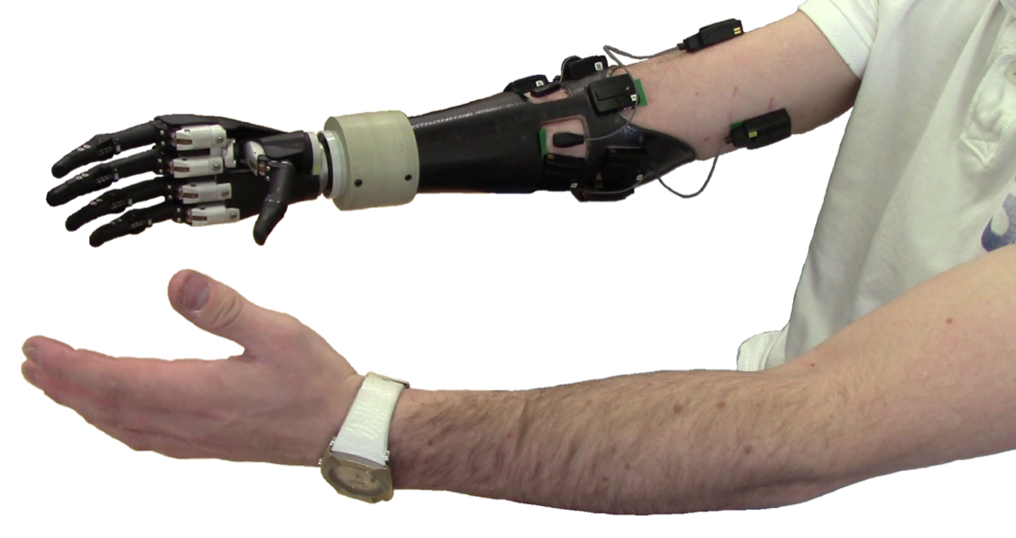
A couple of weeks ago I broke my collarbone. This has meant keeping my arm in a sling for a while, and making do with only one hand. While slightly inconvenient, it’s not the end of the world. In a few weeks I’ll be back to normal operation. It has, however, given me a new understanding of how difficult it is to get things done with only one hand. Previously simple tasks like tying my shoelaces are now impossible!
As I said, for me this will be short lived. For some people though, born missing a limb, or perhaps losing one in an accident, this is something they have to live with day in, day out. As a Biomedical Engineer, my work involves using engineering principles to tackle clinical problems, hopefully improving the quality of life of people with disabilities.
All of which is a rather long-winded introduction to a paper we have recently published on our research to make better artificial (prosthetic) hands.
Computer modelling
The focus of my research has been understanding human movement (in particular involving arms and hands) by building computer models. We use these models to help us understand what goes wrong in certain diseases or injuries, and to design better medical devices and treatments. Our latest paper shows how we might use a computer model of the hand to design a better prosthetic hand.
A better prosthetic hand
Over the last few years, prosthetic hands have become better and better. They now have individually moving fingers and thumbs that are almost as good as the real thing. But there are still a couple of problems. People find it difficult to control many different actions at the same time: they can open or close the fingers, they can bend or rotate the wrist, or they can move the thumb in and out. But not all at the same time, and that makes the use of the hand a bit unnatural. It takes too much effort. Together with our partners at RIC / Northwestern and Cleveland State Universities, we are trying to make a better interface for a prosthetic hand that will make using it really natural.
We have built a computer model of the hand, or ‘virtual hand’, that predicts how the missing hand of an amputee would behave if it were still there. The vision is that this could be used to control an a prosthetic hand worn by an amputee. We would know what the user wanted to do with their hand by recording the signals from the remaining nerves in the arm; the virtual hand would tell us what the prosthetic hand should do. Of course, for this to work, the movements of the ‘virtual’ hand would have to be known as quickly as they would from a real hand. This is known as ‘real-time simulation’.
The real-time hand model showing off postures from the American Sign Language.
The paper describes how the model works, how it simulates the actions of the missing fingers, and how it does it in real time. You can access the full text of the publication here if you want to know how it works (warning: lots of equations!).
Closing the loop
As well as allowing the user to ‘talk to the hand’ (telling the hand what it should be doing), this approach allows the hand to talk to the user! That is, we can simulate the signals from the missing hand that would tell the user where their fingers are, how fast they are moving and whether they are gripping something.
A prosthesis user testing a new type of control for an artificial hand. Image courtesy
of Newcastle University.
This closing the loop of the control system has the potential to really take artificial hands to the next level. This is what the Senseback project aims to achieve and will be the subject of our next publication.
...
Thanks to Dimitra Blana, Wendy Murray, Ton van den Bogert & Kia Nazarpour.
ISTM recently ran a blog writing competition that was open to PhD students and young researchers with a view to improving their lay-writing skills and helping ISTM to play a greater role in the public dissemination of its research. After concluding the competition we will be publishing each entry in turn over the coming months. The 3rd prize winner of our competition was Rachel Gater, a PhD student in the Centre for Doctoral Training, ISTM.
*************************************************
(Source: Microsoft word - Clip Art)
The retina is a complex layer of tissue at the back of the eye. It turns the visual information entering the eye into electrical signals, which are then sent to the brain for processing. Eye diseases such as glaucoma and macular degeneration can damage the retina, leading to significant sight problems and even blindness if left untreated. Glaucoma causes damage to the retina by a build up of pressure inside the eye, whilst macular degeneration involves deterioration of the centre of the retina; the most sensitive region. Although there are some existing treatments for eye diseases such as intravitreal injections and surgery, the success rate of these treatments is not very high and they can cause unpleasant side effects. Therefore, scientists in the field of regenerative medicine are working to develop better treatments for eye diseases.
There are generally two methods that scientists in the field of regenerative medicine can try when researching new treatments for eye diseases. The first approach is the possibility of triggering self repair processes (endogenous regeneration) to help the damaged eye tissues repair themselves. Certain amphibians, such as the newt, are already naturally able to replace their entire eye through endogenous regeneration! We still don’t fully know how amphibians do this, but it is believed that they store stem cells in certain areas of the eye. Stem cells are special because they have not yet transformed into a specific cell type and can therefore be triggered to turn into any type of cell. Therefore if the amphibian’s eye gets injured, the stored stem cells can replace any damaged cells and repair the eye. If scientists can work out the biology of exactly how amphibians do this, then they may be able to help trigger the same process for humans in the future!
(Source: Microsoft word - Clip Art)
Figure 1: Amphibians such as the newt are already naturally able to replace their entire eye through self repair processes (endogenous regeneration).
The second approach scientists in the field of regenerative medicine can try is the possibility of repairing damaged eye tissues using stem cell therapies. Stem cell therapies can be generated from sources such as embryonic stem cells (ESCs), induced pluripotent stem cells (iPSCs) and mesenchymal stem cells (MSCs). As shown in Figure 2, the retina of the eye is made up of many layers each containing different types of cells. These cells include rods (which help us with vision in darkness at night time) and cones (which help us with colour vision in the day time). There are also bipolar cells which do a bit more processing in the nuclear + plexiform layers, as well as ganglion cells which are important in helping to pass on information to the brain. As we know that stem cells can be triggered to turn into any type of cell, scientists believe that we might be able to turn stem cells into new retina cells such as rods, cones and ganglion cells. If successful, these cells could then be used to repair the retina if the eye gets damaged by disease or injury.
(Source: original)
Figure 2: The retina is made up of many layers each containing different types of cell.
So far scientists have been able to turn stem cells into rods, cones and ganglion cells inside a cell culture plate, but there is more work to do before the method can become a treatment given to patients. Even though we can grow the cells, one of the first challenges is figuring out how we can safely deliver the treatment into the patient’s eye. As the eye is so small and fragile, injection needles or surgery may cause further damage to the eye. Therefore, it is important to figure out the best way to precisely deliver the cells to the eye without it causing further damage or being painful for the patient. A second challenge is getting the cells to integrate and function correctly once they are inside the eye. Even if we can deliver the cells correctly, we would need to make sure that the cells are alive, in the correct place and performing their correct function once inside. For example, if we manage to deliver new cone cells into the eye and they correctly perform their role in colour vision, then we would know that the treatment has worked! Finally, a third challenge scientists will need to overcome is the possibility of immune-rejection. As stem cell therapies don’t always use the patient’s own cells, there is a chance that transplanted cells may be rejected, in the same way that a heart transplant might be rejected after heart transplantation surgery. To overcome this challenge, drug treatments such as immuno-suppression therapy may be used to help reduce the risk of rejection. Alternatively scientists might be able to use a patient’s own stem cells which would not be rejected by the body.
In summary, due to the low success rate of current treatments, scientists in the field of regenerative medicine are working to develop better treatments for eye diseases. The two main methods for this include the possibility of triggering self repair (endogenous regeneration) like amphibians can do naturally, or the use stem cell therapies to repair damaged eye tissues. Although scientists can turn stem cells into retina cells relatively successfully in a cell culture plate, challenges still need to be overcome. These challenges include figuring out how to safely deliver cell therapies into the eye, getting transplanted cells to function correctly once inside the eye and overcoming the risk of immune-rejection. If scientists can successfully overcome these challenges, these approaches are likely to transform the way that we treat eye diseases in the future!
Written by Rachel Gater, PhD Student, Centre for Doctoral Training, ISTM (3rd Prize in the ISTM Blog Post Competition 2016)
Research led by ISTM and published this week in Diabetologia (the journal of the European Association for the Study of Diabetes) has identified a new link between pre-eclampsia in pregnancy and the development of diabetes in later life.
The condition, which results in high blood pressure and protein in the mothers’ urine, affects 5-8% of pregnancies and is the most common cause of severe perinatal ill health. The study showed that pre-eclampsia is independently associated with a two-fold increase in future diabetes. This increased risk occurs from less than 1 year after delivery of the baby and persisted to over 10 years after birth.
Dr Pensee Wu, ISTM
Dr Pensee Wu, Lecturer in Obstetrics and Gynaecology at ISTM, is the first author of this publication and said:
“This study highlights the importance of clinical risk assessment and follow-up for the future development of diabetes in women with pre-eclampsia. Understanding of health conditions during pregnancy and their impact on health over a woman’s life is vital in the prevention of conditions such as diabetes.
“Ensuring women are screened regularly and take preventative measures through diet and exercise could help reduce the number of women who later contract diabetes after experiencing pre-eclampsia during pregnancy.”
The study involved a systematic review of research over the past 10 years, much of which was conflicting about the impact of pre-eclampsia later in life.
The understanding of the long term impact of women’s health following pre-eclampsia is however growing. The American Heart Association has linked pre-eclampsia to longer term cardiac conditions.
“Diabetes is a multi-organ condition. If we can prevent it from developing early on, it could dramatically reduce the risks of serious health issues later in life for women after birth” says Dr Wu.
Researchers hope that dissemination of this study to clinicians, particularly those in Primary Care health provision, will inform practice and longer term preventative measures.
As well as being a Lecturer in Obstetrics and Gynaecology at ISTM, Dr Pensee Wu is also an Honorary Consultant Obstetrician and Fetal Medicine Subspecialist at the University Hospital of North Midlands (UHNM).
The latest issue of the ISTM Translate magazine is now available in digital and print format. The theme focus for this issue is nanopharmaceutics.
There are contributions from Dr Clare Hoskins, Dr Anthony Curtis, as well as other members of the Keele Nanopharmaceutics Group. Articles include...
· Medicine at the small scale: The exponential growth in nanopharmaceutics
· Programming death in cancer cells: Novel technologies improving long-term prognosis
· The great solubility challenge: Investigating potential in targeted drug delivery
· Theranostic Advances: The knowledge and fabrication of nanotechnologies
· Nanopharmaceutics Symposium: Successful second annual event at Keele University
· Spotlight: The people behind ISTM
· Public engagement: Bringing research closer to people
· Taking advantage of change: A new streamlined Institute structure for ISTM
By clicking here you can access the online digital version of Translate Issue 4. Alternatively you can request a paper copy by contacting: Joseph Clarke +44 (0)1782 674998 | j.clarke@keele.ac.uk
Written by Dr David Mottershead (Lecturer in Biochemistry & Cell Biology, ISTM) ********************************
Making new friends from the Congress. (David is second from right).
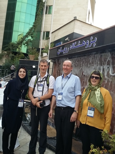
As a result of an email I received on the 20th of June this year inviting me to speak at the Royan International Twin Congress (August 31-September 2) in Tehran, Iran, I was plunged into quite an adventure. The Royan International Twin Congress has been held every year since the first meeting in September 2000. This year’s meeting saw the joint holding of the 17th Congress on Reproductive Biomedicine and the 12th Congress on Stem Cell Biology & Technology, both supported and organized by the Royan Institute in Tehran. This being my first trip to the middle east meant that a new and unusual experience was assured.
Ceiling of the Tomb of Hafez, Shiraz.
The meeting itself reflects the nature and makeup of the Royan (in Persian “royan” means embryo) Institute itself, which has Divisions devoted to reproductive biomedicine, stem cell biology and biotechnology. The congress itself has the two streams of reproductive biomedicine and stem cell biology running concurrently. This enabled one to pick and choose between both session streams, and although I found myself predominantly within the reproductive stream, I did cross over for a number of very interesting presentations within the stem cell stream as well. International speakers from across the world were in attendance (UK, USA, Austria, China, Netherlands, Australia, Germany, Denmark, Sweden, Switzerland, Italy, for example). Apart from Keynote Lectures and Award Lectures, there were the various invited speakers talks, short oral presentations, poster sessions and exhibitions. The sessions of particular interest to me were, Animal Biotechnology, Tissue Engineering in Reproductive Sciences, and Regenerative Medicine, Novel Discoveries.
Tombs of kings past, Naqsh-e-Rostam.
The conference organizers looked after the invited speakers extremely well, we were never short of food/refreshments at the venue and every evening we were taken to see sites in Tehran and out to eat, enabling us to experience the magnificent local cuisine. From my own point of view, the congress was extremely productive with good discussions/connections established between myself and two separate labs at the Royan Institute which should lead to ongoing collaborations. For all the invited speakers a 2 day tour of highlights of Iran was offered which I participated in, enabling one to experience the history of the country and see some amazing sites. First we flew South to Shiraz, a distance of 700 km. We arrived late and would not have time to get to know Shiraz well, but did visit the Tomb of Hafez, one of Iran’s famous and revered poets who lived in the 14th century (1325-1389). Upon leaving Shiraz by bus we started our long trip back North to Tehran. Our first stop 70 kms from Shiraz was at a series of tombs cut into the cliff side where ancient kings were buried. The site is known as Naqsh-e Rostam and had a distinctly “Indiana Jones” feel to it, certainly it transported one back in time. Not far from this site we stopped at the ruins of the ancient city of Persepolis which was the ceremonial capital of the Achaemenid Empire (550–330 BC). Here various kings and statesmen from around the world would come to greet the Iranian king, a must see site on any trip to Iran. Finally we arrived at the city of Esfahan (also spelt Isfahan) about 300 km South of Tehran. Esfahan is the 3rd largest city in Iran at nearly 2 million people. The main attraction is the large Naghsh-e-Jahan square constructed between 1598 and 1629. Around the square are located three mosques and what seems like hundreds of shops selling the most beautiful craftworks. I could not do this area justice in this short visit, however, given the collaborative links which I believe will be established, it may well not be my last.
One of the mosques at Naghsh-e-Jahan Square, Esfahan.
I was surprised and delighted to find a couple of weeks ago that I had been awarded the Vice-Chancellor’s PhD Prize 2016 for the best postgraduate research at the University of Greenwich.
My programme of research was Understanding MS/MS Fragmentation of Small Molecular Weight Molecules. I proved the hypothesis that protonation altered the conformation of molecules such that some bonds were weakened and more susceptible to cleavage. This impacts all scientists who use MS/MS for structural elucidation in that quantum chemistry can now be used a tool in assignment of ion structures. I am pleased to have been able to make a real difference to UK science.
I loved every minute of my PhD, particularly the stimulating scientific discussions with my supervisor Professor Frank Pullen and computational chemistry expert Alexander Alex. I also enjoyed getting to grips with quantum chemistry.
Thanks to the support and encouragement of the staff in the Greenwich University Department of Pharmaceutical, Chemical and Environmental Sciences, I managed to take my viva voce one day before the third anniversary of starting my research programme, passing with no changes required to my thesis and having published five research papers and two editorials. I am also grateful for the tolerance of my family who put up with me working all the time!
Patricia has since come to work at Keele University for ISTM in the breath analysis research group pioneered by Professor David Smith FRS.
Friday 2 September 2016
How brain implants can let paralysed people move again
Dimitra Blana, Keele University and Andrew Jackson, Newcastle University
Something as simple as picking up a cup of tea requires an awful lot of action from your body. Your arm muscles fire to move your arm towards the cup. Your finger muscles fire to open your hand then bend your fingers around the handle. Your shoulder muscles keep your arm from popping out of your shoulder and your core muscles make sure you don’t tip over because of the extra weight of the cup. All these muscles have to fire in a precise and coordinated manner, and yet your only conscious effort is the thought: “I know: tea!”
This is why enabling a paralysed limb to move again is so difficult. Most paralysed muscles can still work, but their communication with the brain has been lost, so they are not receiving instructions to fire. We can’t yet repair damage to the spinal cord so one solution is to bypass it and provide the instructions to the muscles artificially. And thanks to the development of technology for reading and interpreting brain activity, these instructions could one day come direct from a patient’s mind.
We can make paralysed muscles fire by stimulating them with electrodes placed inside the muscles or around the nerves that supply them, a technique known as functional electrical stimulation (FES). As well as helping paralysed people move, it is also used to restore bladder function, produce effective coughing and provide pain relief. It is a fascinating technology that can make a big difference to the lives of people with spinal cord injury.
Dimitra Blana and her colleagues at Keele are working on how to match this technology with the complex set of instructions needed to operate an arm. If you want to pick up that cup of tea, which muscles need to fire, when and by how much? The firing instructions are complicated, and not just because of the large number of core, shoulder, arm and finger muscles involved. As you slowly drink your tea, those instructions change, because the weight of the cup changes. To do something different, like scratch your nose, the instructions are completely different.
Instead of just trying out various firing patterns on the paralysed muscles in the hope of finding one that works, you can use computer models of the musculoskeletal system to calculate them. These models are mathematical descriptions of how muscles, bones and joints act and interact during movement. In the simulations, you can make muscles stronger or weaker, “paralysed” or “externally stimulated”. You can test different firing patterns quickly and safely, and you can make the models pick up their tea cups over and over again – sometimes more successfully than others.
Modelling the muscles
To test the technology, the team at Keele is working with the Cleveland FES Center in the US, where they implant up to 24 electrodes into the muscles and nerves of research participants. They use modelling to decide where to place the electrodes because there are more paralysed muscles than electrodes in current FES systems.
If you have to choose, is it better to stimulate the subscapularis or the supraspinatus? If you stimulate the axillary nerve, should you place the electrode before or after the branch to the teres minor? To answer these difficult questions, they run simulations with different sets of electrodes and choose the one that allows the computer models to make the most effective movements.
Currently, the team is working on the shoulder, which is stabilised by a group of muscles called the rotator cuff. If you get the firing instructions for the arm wrong, it might reach for the soup spoon instead of the butter knife. If you get the instructions to the rotator cuff wrong, the arm might pop out of the shoulder. It is not a good look for the computer models, but they don’t complain. Research participants would be less forgiving.
Knowing how to activate paralysed muscles to produce useful movements like grasping is only half of the problem. We also need to know when to activate the muscles, for example when the user wants to pick up an object. One possibility is to read this information directly from the brain. Recently, researchers in the US used an implant to listen to individual cells in the brain of a paralysed individual. Because different movements are associated with different patterns of brain activity, the participant was able to select one of six pre-programmed movements that were then generated by stimulation of hand muscles.
Reading the brain
This was an exciting step forward for the field of neural prosthetics, but many challenges remain. Ideally brain implants need to last for many decades – currently it is difficult to record the same signals even over several weeks so these systems need to be recalibrated regularly. Using new implant designs or different brain signals may improve long-term stability.
Also, implants listen only to a small proportion of the millions of cells that control our limbs, so the range of movements that can be read out is limited. However, brain control of robotic limbs with multiple degrees-of-freedom (movement, rotation and grasping) has been achieved and the capabilities of this technology are advancing rapidly.
Finally, the smooth, effortless movements that we usually take for granted are guided by rich sensory feedback that tells us where our arms are in space and when our fingertips are touching objects. However, these signals can also be lost after injury so researchers are working on brain implants that may one day restore sensation as well as movement.
Some scientists are speculating that brain-reading technology could help able-bodied individuals to communicate more efficiently with computers, mobile phones and even directly to other brains. However, this remains the realm of science fiction whereas brain control for medical applications is rapidly becoming clinical reality. Dimitra Blana, Research Fellow in Biomedical Engineering, Keele University and Andrew Jackson, Wellcome Trust Senior Research Fellow, Newcastle University
This article was originally published on The Conversation. Read the original article.
ISTM recently ran a blog writing competition that was open to PhD students and young researchers with a view to improving their lay-writing skills and helping ISTM to play a greater role in the public dissemination of its research. After concluding the competition we will be publishing each entry in turn over the coming months. The 2nd prize winner of our competition was Homayemem Weli, a PhD student in Cell and Tissue Engineering at ISTM.
*************************************************
Walking through the streets of London in mid-summer, I couldn’t help but notice its beautiful 'ornaments' of modern architecture, such as the "Gherkin" or the "Walkie-Talkie". Imposing as these are, they attract less curiosity than Wiltshire’s Stonehenge.
A graceful and strong modern building speaks of a firm foundation and good design, but an ancient monument raises questions such as "when", "how" and "why". Some of us recall the creation of the London skyscrapers - having possibly witnessed it - but I dare say none of us witnessed the making of Stonehenge! Instead, we rely on archaeological research to answer questions about its origin, age and significance. This ‘discovery science’ compels us to find out why things are the way they are, and go from the known to the unknown.
Stonehenge by David Ball -
www.davidball.net
Keen to unravel the unknown, I set out to study an aspect of why people age. My focus was on women and what happens to their vaginal and skin tissues before and after pregnancy. Adorned with laboratory clothing and gloves, as though performing carbon dating on the standing stones of Stonehenge, I examined the structure of collagen fibres and cross-links within the tissues. Collagen is a protein that supports structures within the body. Cross-links are bonds formed between collagen groups (called fibres) or between chemical substances such as amino acids or reducing sugars. The larger the number of certain cross-links within the collagen fibres, the older the fibres. Collagen 'cross-link dating' can separate old fibres from young ones.
I tested two groups of tissues, pregnant and non-pregnant, of similar biological ages. I separated a particular cross-link, pentosidine, which is a known marker of tissue ageing, from the tissue solutions with liquid chromatography (a method for identifying and separating substances present within a solution). I noted the amount of pentosidine in each tissue, and compared the values.
This process of 'cross-link dating' separated the age-matched tissues into two groups – ‘old’ and ‘young’. Before pregnancy, tissues were ‘older’, but after pregnancy, they appeared ‘younger’. During pregnancy, the signs of ageing appeared to reverse!
Studying further, I discovered this was linked with a rise of a potent antioxidant, glyoxalase I, in the tissues during pregnancy. Antioxidants such as glyoxalase I protect the body cells from molecules that could cause damage or promote ageing.
Antioxidant glyoxalase I enzyme
expression in vaginal tissues during (left) and after (right) pregnancy. Green
glow, clearly visible within the pregnant tissue, represents presence of the
antioxidant enzyme. The pregnant tissues had more antioxidant.
Oestrogen, a well-known pregnancy hormone, influenced the amount of the antioxidant in the tissues. I found higher oestrogen levels in pregnant tissues as shown in the images below. This implied pregnancy resulted in higher oestrogen and antioxidant levels.
Oestrogen receptor expression in vaginal
tissues during (left) and after (right) pregnancy. Red dots signify oestrogen
activity within the tissue and show raised level of oestrogen during pregnancy.
I concluded that oestrogen influences the ‘age’ of collagen fibres of the skin and vaginal wall by increasing the antioxidant glyoxalase I. Rise in oestrogen as seen in pregnancy leads to rise in the enzyme, subsequently retarding collagen fibre ageing within the tissues. In this way, pregnancy results in younger appearing tissues.
Oestrogen is a female reproductive hormone that changes throughout the life of a woman. It increases in quantity during pregnancy and reduces as women grow older, finally reaching its lowest levels in menopause. My finding shows that pregnancy may retard this ageing process in the vaginal and skin tissues of women. A previous study noted a reduction in similar ageing cross-links within blood vessels also in association with higher oestrogen levels, suggesting that this effect may exist in many body tissues.
By studying pregnancy, I discovered a relationship between oestrogen, an antioxidant, and the ‘age’ of collagen fibres (change of the structure) in skin and vaginal tissues.
New knowledge can be gained from investigating age-old body processes. It's always worth asking "when", "how" and "why”!
Written by Homayemem Weli, PhD Student, ISTM (2nd Prize in the ISTM Blog Post Competition 2016)
This is a meeting for people who work in the area of shoulder biomechanics. We try to understand how the shoulder works, what goes wrong after injury or disease, how to improve its movement in sport, and how to protect it in well-designed work environments. For those of us doing shoulder research (and probably nobody other than us) it is a fascinating topic, and we look forward to getting together every couple of years and discussing it.
My particular focus is mathematical modelling of the shoulder. Using models allows us to investigate how muscles and joints work without invasive procedures on actual people. It is a very useful tool, and one of the ways we use it is to design technological systems that tackle paralysis.
Co-presenter Ricardo Matias (University of Lisbon) during the modelling workshop
As part of the International Shoulder Group meeting this year, I ran a modelling workshop that aimed to introduce modelling to people who have not used it before. The response was very positive, and everyone seemed keen to give it a go. With participants from 13 different countries, it is great to know that our models could be used all over the world!
Dimitra and her thank-you gift for helping to organise the conference
Shoulder biomechanists are such a lovely lot, and I can’t wait to see everyone again in 2018!
ISTM recently ran a blog writing competition that was open to PhD students and young researchers with a view to improving their lay-writing skills and helping ISTM to play a greater role in the public dissemination of its research. After concluding the competition we will be publishing each entry in turn over the coming months. The 1st prize winner of our competition was Fraser Philp a PhD student at ISTM. Fraser's PhD focuses on identifying within current practice and research, methods used for predicting injury and performance within football and Fraser used this topic as the theme for his blog post.
*************************************************
Association football or soccer is one of the most popular international sports, with approximately 200,000 professionals and a further 240 million amateur male and female participants worldwide. Football is England’s largest national team sport, with men’s and women’s football being the first and third largest team sports respectively. Associated with the high levels of participation in football is a high level of injury risk. As many as 47% of footballers have been forced to retire from the game due to injury throughout the season, an average outfield player is expected to sustain at least 1-2 injuries resulting in them being unavailable for 1 competitive game. High rates of injury can negatively impact on the performance of an individual. Likewise an increased number of individuals sustaining injury within a team can negatively affect team performance, which, in a competitive league can have further consequences.
Given the problems associated with injury, the medical and sports science teams who work with professional football teams try to minimise injuries occurring. One of the ways they attempt to do this is through screening. Screening can involve exercise tests and measures of physical performance that are used in an attempt to identify injury risk factors. These tests are usually carried out before the competitive season starts, in a period known as pre-season, and during the competitive season itself. Despite the widespread use of these tests and measures, many of them have not been and compared against other methods of measurement for validation purposes.
Motion capture is more widely known for its use in the movie industry in virtual recreation. It is also used as the gold standard measurement in hospital settings for measuring walking patterns and human movement patterns in people with neurological disorders. In order to measure the movements of the FMS with motion capture cameras, some additional preparation is needed. This requires placing reflective markers on selected body parts of the person. This is because the motion capture camera’s only pick up reflections from the infrared light that they send out. These markers can then be virtually recreated providing an outline of the person’s body parts on which they were placed. Once this has been done we are able to see the angles achieved by the participant in all 3 dimensions i.e. how much they bent their knee or how much their hip was rotating. It also allows for a description of the movement patterns that are occurring across the joints when the participant completes the FMS. Furthermore we can also attribute numerical values to the rules and scoring criteria of the FMS exercise tests.
Figure above shows the process after placing the markers on and then virtualy recreating them.
Alongside this we have monitored a football team over one competitive season and will investigate whether there is a link between the measurements we took and the injuries they sustained. Within this analysis we will also be investigating things such as the amount they trained, the surface they trained on and what their match fixtures were like. Hopefully a better understanding of all these factors will allow for fewer injuries in footballers.
Written by Fraser Philp, PhD Student, ISTM (1st Prize in the ISTM Blog Post Competition 2016)
Professor Christine Roffe, Consultant in Stroke Medicine at the Royal Stoke University Hospital and Lead of the Stroke Research in Stoke Group at ISTM, has been awarded a major grant to fund a £2.5 million project from the NHS National Institute for Health Research (NIHR) Health Technology Assessment Programme for her MAPS-2 clinical trial.
Professor Roffe has a proven track record in shaping the development of acute and rehabilitation stroke services in North Staffordshire, with research that has gone on to influence regional and national practices in stroke care. As Principle Investigator, Professor Roffe will lead a consortium which includes Keele University in collaboration with the University Hospitals of North Midlands (UHNM), the University of Birmingham and Anglia Ruskin University. The consortium will investigate methods to reduce pneumonia in stroke patients.
Pneumonia is a major cause of death and disability after stroke. Stroke patients are at risk of contracting pneumonia through the inhalation of saliva or gastric content after vomiting or regurgitation which contain harmful bacteria. This award from the NIHR will fund clinical trials that will test two methods for preventing pneumonia. One will aim to prevent patients vomiting in the first place, while the other will use an antibacterial paste in the patient’s mouth to reduce the harmful bacteria in their saliva.
Professor Roffe said that: “Pneumonia after stroke weakens patients and delays recovery. In this trial we are hoping to show that these treatments not only prevent pneumonia and death, but also allow patients to recover better and faster.”
The trial will begin to recruit patients in October 2016 and run for three years. If the treatments prove effective, lives could be saved and stroke patients may stand a better chance of getting back to their normal life in a shorter space of time.
I’m A Scientist Get Me Out Of Here is a competition in the vein of I’m A Celebrity Get Me Out Of Here, where several people compete for a prize and each week, one of them is evicted until only one remains. Luckily ISTM's Matt Dunn didn’t need to go to the jungle as I’m A Scientist takes place entirely online at http://imascientist.org.uk/ . Beating off fierce competion, Matt went on to win the competition. So, we asked Matt to tell us a little bit more about his experience...
The competition ran from 13th to 24th June, where scientists with successful applications were split into different ‘Zones’ depending on their field, I was put into the ‘Cells Zone’ with four other cell biologists. For the next two weeks, we took part in numerous 30 minute live-chat sessions with classes of students from twenty different secondary schools, ranging from Year 7 to Year 9, all across the country. In these live chats the students could ask us anything they liked, from the work we did, to describing an average day, to what our favourite foods were! Despite the casual nature of the chats, the students can really test your knowledge of your subject, making it a great challenge.
Outside of the live chats, students can ask you questions directly on a profile, and they vote for their favourite scientist. Scientists with the least votes were evicted every day for the last five days until only one was left. I was lucky enough to be the last scientist standing, winning £500 to spend on outreach activities!
I plan to use the money to raise awareness of the great regenerative medicine work we all do here at ISTM, as part of the HEART Outreach group. We usually attend a few fairs and events a year as exhibitors, to spread the good word, but with the prize money we will be able to travel further and better show off the quality of the work we do here with improved cutting-edge technology.
I’m A Scientist runs multiple times a year, so I’d recommend it for anyone interested in outreach and science communication, as what is the point of doing the work we do if we can’t explain it to school children?
We are keen to encourage more of our young researchers and students to engage with our ISTM Translate blog and are keen for you to play a bigger role in writing and editing articles for it. The blog has global reach with well over 5000 views to date and regular visitors from across 10 different countries. As well as having the opportunity to promote the Institute on an international level and to communicate stories and information that are of interest to you to a wider audience, you also have the chance to hone your lay-writing skills.
As you may have already heard, Keele University is now a member of The Conversation. The Conversation is an independent source of news and views, sourced from the academic and research community and delivered direct to the public. Their team of professional editors work with researchers to unlock their knowledge for use by the wider public.
So with a view to improving your lay-writing skills and helping ISTM to play a greater role in the public dissemination of research, we have decided to hold a competition to which you are all invited to participate.
Please see the information below from Dr Dimitra Blana, who will be helping to facilitate this writing competition...
You are invited to submit a post to the ISTM Translate blog describing a research topic of your choice to the general public.
Why, you ask? Because it will help you improve your public engagement skills, which is increasingly important for scientists. If that's not enough, there is also a prize, so read on!
Your blog post should be a lay-term summary of either a research paper you recently published, or the scientific area you are investigating. It should be no more than 1000 words and include some compelling images.
Submit your blog post and images to me over email (d.blana@keele.ac.uk) by 30th June to be entered into a competition to win a cash prize. First prize is £100 and the Runner Up prize is £50! The winners will be announced during a lunch party in the Guy Hilton Research Centre on Thursday 21st July.
A few points to consider when preparing your post:
Make sure the language is appropriate for the general public. To ensure that everyone can understand your writing, it will be judged by people outside your research area. I am thinking of enlisting my 14 year old Greek cousin.
Journal paper writing rules do not apply: be natural and informal. Of course make sure that the science is sound!
1000 words is the upper limit, but try to keep it short and engaging. Can you keep my 14 year old Greek cousin's interest for 1000 words? Or will she switch to cat videos half way through?
Use images you have permission to use. And remember that journal paper result figures are not usually considered compelling by the general public.
Dr Paul Roach (Left) and Dr Ed Chadwick (Right) introduce themselves.
Last week, ISTM held a successful open afternoon for people interested in studying a post-graduate degree in Medical Engineering. The Open afternoon attracted a good intake of participants from a range of different backgrounds. The participants were able to learn about the School of Medicine's MSc courses in Biomedical Engineering and Cell and Tissue Engineering, and learnt about the cutting edge research being carried out by the Institute for Science and Technology in Medicine. They also had the chance to see state of the art laboratories and talk to current and former students and researchers.
Dr Ed Chadwick, who organised the event along with Dr Paul Roach, said "We were delighted to see such strong interest in our research and the courses we offer from the Keele undergraduate community and those from the wider region that were able to join us. People often have a fairly vague idea of what medical engineering comprises, and it was really nice to see that they were genuinely interested and indeed excited by what we do. The people who work in the area are typically from really diverse academic backgrounds, and this was reflected in the people attending. The day was definitely a success with really strong interest, and we look forward to doing it again!"
This prestigious award is funded by the Wellcome Trust as part of an Academy of Medical Sciences scheme to promote strategic research links and scientific collaborations between the UK and the Middle East.
Dr Adams will build upon a recent collaboration between the Chari-Lellouche groups, which aims to develop nanotechnology based tools for safe genetic engineering and non-invasive imaging of neural transplant cells.
Professors Alicia El Haj (ISTM) and Tsachi Keren-Paz (Law) hosted the second seminar in the ESRC series 'Liability versus innovation: unpacking key connections'.
The seminar brought together the Vice-Chancellor, clinicians, plaintiff personal injury lawyers, a Medical Defence Union representative, health economists, ethicists (Profesor Wendy Rogers, Macquarie University, Sydney) and academic lawyers from the United States (Professor Alex Stein, Benjamin N. Cardozo, School of Law, NY, Australia (Associate Professor Tina Cockburn, QUT Law School, Brisbane) and the UK.
Discussions revolved around the questions whether the threat of tort liability stifles innovation, the extent to which the different liability rules in the USA, UK and Australia stifle to a different extent innovation, and the methodological question how to define and measure innovation and how to measure the effect of legal rules on levels of innovation.
Are you are interested in a post-graduate degree in Medical Engineering?
If so, please join us to find out what we have to offer. You will learn about our MSc courses in Biomedical Engineering and Cell and Tissue Engineering, and hear about the cutting edge research being carried out by the Institute for Science and Technology in Medicine. You will have the chance to see state of the art laboratories and talk to current and former students and researchers.
The Open Afternoon will take place at the Guy Hilton Research Centre from 13:30 - 16:30 on May 4th 2016. To confirm your attendance, please register by leaving your details below (by the 1st of May). Transport will be provided from Keele University campus.
The Director of ISTM & members of ISTM pose with the Tongji delegation
ISTM was delighted to welcome a delegation from Tongji University, Shanghai, China last week on the 5th April.
Dean of Medicine, Professor Xu Guotong; Professor Peng Luying; Professor Liang Xingqun; and the Director of the International Office, Associate Professor Zheng Hao met with members of ISTM as well as colleagues from across the University to discuss future research collaboration in the areas of Medicine.
The delegation, part of a larger delegation that also included experts in Environmental Sustainability, also visited the School of Medicine and the Royal Stoke University Hospital. The delegation was hosted by the Vice Chancellor, Professor Trevor McMillan and the visit to Keele University culminated in the signing of a Memorandum of Understanding between the two universities.
Tongji University is part of the prestigious 985 group of universities selected by the Chinese government for special support and has one of the country's highest ranked medical schools. In 2015 Tongji and Keele successfully applied for joint ERASMUS+ funding for staff mobility and incoming PGR mobility.
Congratulations to the following members of ISTM on their recent promotions...
Ed Chadwick moved to Keele as Lecturer in Biomedical Engineering in 2012 from Aberystwyth University where he taught Biomechanics. After obtaining a degree in Mechanical Engineering at Nottingham and PhD in Bioengineering from Strathclyde, Ed spent several years working as a Senior Research Associate at Case Western Reserve University in Cleveland, Ohio and at Delft University of Technology in the Netherlands.
His research interests are in the application of biomechanical modeling and simulation techniques to study upper limb function in a range of neuromuscular disorders including spinal cord injury and stroke. He has a particular interest in the use of functional electrical stimulation (FES) and development of FES devices for the restoration of function. He is currently the theme lead for the Rehabilitation Research Group within ISTM, and MSc course Director for Biomedical Engineering, leading modules in medical technology and devices.
As well as a range of PhD studentships, charity and industrial funding, Ed is a co-investigator of a major EPSRC project in Enabling Technologies for Sensory Feedback in next generation assistive devices, in collaboration with Imperial, Newcastle, Leeds, Essex and Southampton universities. He also continues to collaborate with Case Western Reserve University in the USA on a major project funded by the National Institutes of Health. Ed currently serves on the Executive Council of the International Society of Biomechanics, and was one of the recipients of Keele's MRC Centenary Awards.
Clare Hoskins has been promoted to Senior Lecturer in recognition of her research and her contribution to development of the School of Pharmacy. Clare has managed stage 2 of the MPharm programme for the past three years and is currently leading the restructuring of the year-long module in line with the newly accredited course. Clare has also contributed towards the School's recruitment and widening participation activities and sits on the Faculty Outreach Committee.
Clare has taught on all years of the degree and especially is interested in ensuring her teaching content is informed by the advances in research in her field of interest. Clare's research group, "Keele Nanopharmaceutics", has gained significant recognition over the past four years for its work in the use of nanomedicines for drug delivery. Currently Clare supervises seven PhD students who work on multi-disciplinary projects ranging from organic synthesis through to biological investigations. Clare is the elected Secretary of the Royal Society of Chemistry, Chemical Nanosciences & Nanotechnology Division as well as sitting on the Committee for Preclinical Sciences and Animal Health for the Controlled Release Society. Clare organised the Nanopharmaceutics Symposium at Keele in July 2015, which she intends running on an annual basis.
Paul Roach joined Keele in Nov 2009 and has since established a laboratory for microfabrication and surface analysis. Learning from natural biological processes, he has pioneered the understanding of interfacial interactions with nano-surfaces. His work has generated international interest with his publications receiving over 3,300 citations with an H-index of 14.
He has attracted external grants totaling over £600k and is also a co-investigator of the EPSRC-MRC Regenerative Medicine Centre for Doctoral Training in conjunction with Nottingham and Loughborough worth 3.5m overall. In addition, Paul was a key contributor to the EPSRC Capital award for major equipment in ISTM. His research and PhD students investigate development of advanced materials, their manufacture and use in directing biological responses.
Paul has taken a very active role in promoting interdisciplinary research and is a prominent member of the biomaterials and regenerative medicine communities in the UK and Europe, serving as adjunct faculty at the Donders Institute for Brain, Cognition and Behaviour at Radboud University in the Netherlands. In 2014 he was elected to the UK Society of Biomaterials council and in 2015 to the Executive Committee of the RSC Biomaterials group. He is also a member of the EPSRC Early Career Manufacturing Forum.
Having previously served as academic conduct officer in the Medical School, Paul is now Director of MSc in Cell and Tissue Engineering and co-ordinates the summer school for Saudi medical students who have been visiting ISTM for the past five years.
After receiving a petition from staff and students at the Institute for Science & Technology in Medicine, the Institute's Executive Management Committee has decided to commission an 8ft (2m 44cm) statue of Professor Alicia El Haj.
A petition calling for the statue was started by a group of enthusiastic PhD students and Post Doctoral Research Assistants at the Institute. They felt that some form of lasting tribute to the work carried out by Professor Alicia El Haj, the Institute's Director, was needed to immortalise her contribution to science and to the Institute. The petition quickly took off attracting more than 1600 signatures. Overwhelmed by the feeling of support for such a tribute, the Institute's Executive Management Committee debated the request and decided unanimously in favour of accepting it.
When asked about having a larger than life statue built of herself, Professor El Haj replied, "Well, it's about time!"
An artists impression of what the statue might look like in front of the Guy Hilton Research Centre
A local artist, Mr A.P. Fool, has been commissioned to build the statue, which will be cast is bronze and mounted on a marble stone. The statue is to be positioned at the front of the Guy Hilton Research Centre and will be visible to all visitors to the building.
Concerns were raised by the Faculty of Health over the projected costs of project, which is currently estimated at £57,000, but these concerns have since been withdrawn based on the proposed funding plan to be implemented by the Institute. Essentially a levy will be placed against all members of ISTM. Known as the "T Account Tax", each member will contribute a proportional amount from their personal Teaching Accounts. By and large though, the response to this arbitrary funding method has been overwhelmingly positive - a tribute in itself to the popularity of Professor El Haj.
The statue is due to be completed, in place and greeting visitors, by the beginning of September this year.
George, was awarded second prize in the Eureka category of the Science Photo Competition 2015, by the EPSRC (www.epsrc.ac.uk/photocomp)
Called 'Curious Neurons' the image shows developing brain cells from the rat cortex, cultured at Keele (ISTM). The cell nuclei are labeled in blue. The red cells are glia, the brain cells that provide structure and support to neurons, the functional component of the nervous system. In this image the neurons, in green, are "feeling" their surroundings, i.e. looking for other cells to connect with (form synapses), so that they can "talk" with each other. This research aims to help identify the environmental elements necessary to grow stem cells for use in therapeutic treatments for people with Parkinson’s and other neurodegenerative diseases. The image was taken at Keele with a Nikon 80i microscope with a Hamamatsu ORCA camera.
One of the judges, Professor Robert Winston, said: “It is crucial to promote greater understanding of science and engineering research, the role it plays in making new discoveries, developing new technologies and in making the world a better place for us all. These are truly inspirational images and tell great stories. It was a real pleasure to take part as a judge and I hope people will want to find out more.”
Sue Hunter and her team have now finished recruiting participants to the NIHR/MRC funded FAST-INdICATE trial, a multi-site Randomised Controlled Trial (RCT) of functional strength training for the arm and hand after stroke; They have successfully met their target of 288 participants across the 3 sites (Norfolk, Birmingham and North Staffordshire) by the end of January 2016, which was the deadline for recruitment. Their participants will still undergo follow-up assessments and this will continue until June/July 2016. The team have been complimented as a site by the trial manager on their consistent level of recruitment and on the quality of their data/records - so they have every reason to be proud of themselves. Data is still being collected and processed in preparation for analysis. Sue Hunt the Principle Investigator for the North Staffordshire site said "Thank you to everyone who has been or continues to be part of this team - all the hard work is very much appreciated!" Further details about the trial can be found on the team's website: www.fastindicate.com
At ISTM we are fully committed to equal opportunities in the workplace. We have a strong track record in challenging the traditional male orientated stereotypes of lab work and in breaking down the barriers that women in science often face. We have a range of procedures and initiatives to help research staff, regardless of gender, advance within their profession by providing them with the skills and opportunities they need to develop.
Having been awarded the Athena SWAN Bronze award in April 2013 in partnership with Keele’s School of Medicine, we are now making headway in pushing forward with initiates previously outline in the Action Plan. With the continuous support from both our male and female members of staff, progress is being made towards our plans to apply for an Athena SWAN Silver award within the next year.
We were also delighted to hear that the School of Medicine has recently been awarded a Daphne Jackson Fellowship, which has allowed Dr Vijayalatha Venugopalan, under the supervision of ISTM’s Professor Paul Horrocks, to restart her career in research after a break of more than two years.
So, to mark International Women’s Day this year and to demonstrate our commitment to gender equality, we asked some of our top female researchers...
What does it take for women to succeed in science?
Professor Alicia El Haj Institute Director for ISTM Professor of Cell Engineering Professor El Haj is a leading figure in Regenerative Medicine and has been involved in bringing together interdisciplinary groups within biomedicine, physical sciences and engineering interested in aspects of cell and tissue engineering.
"I believe that the most important factor to survive in science for both women and men is a passion for seeking answers to the unknown. Most scientists I know are drawn to the field because of the fascination of being able to answer questions in biology or physics or chemistry which underpin the fundamentals of life and can forge a path to new innovations.
As women, we need this same passion. Especially since the lifestyle can be demanding and challenging in terms of commitments, travel and hard work. I also believe a healthy work/life balance is fundamental to any successful career. There are times when we need to be able to switch off and enjoy life, whether that be through family, friends or in solitary.
Finally, and perhaps most importantly, it is important to maintain a sense of humour and a positive attitude, come what may. There are many times when our paths can be thwarted for a multitude of reasons, not least experiments which fail or findings and theories which are criticised. Being able to define a new path with a smile is a very useful trait for maintaining a long and productive career in science."
Professor Fricker leads a research group within ISTM. The Group's goals are to develop treatments for Parkinson’s disease and Huntington’s disease.
"I believe that women need courage, self-belief and resilience to succeed in science. As I’ve progressed through my career I’ve seen how tough it is for scientists to really succeed, whatever their gender. We have limited opportunities, and have to be competitive to flourish. Too often women do not promote their qualities or put themselves forward; we tend to be quieter or less assertive, so that achievements often go unnoticed and unrewarded.
I believe that female scientists need to have more courage to showcase their abilities, and more belief in the value that they bring to research. We need to be more assertive, and to have the confidence to get involved and take on new challenges. I think too that women need the resilience and energy to juggle an often complex work-life balance and to retain a focus on their career path, despite the diversions that may appear."
Professor Christine Roffe Consultant in Stroke Medicine Reader and Honorary Senior Research Fellow Professor Roffe has shaped the development of the acute and rehabilitation stroke services in North Staffordshire, with positive impact on regional and national practice.
"As with every scientist the most important prerequisite is scientific curiosity and the will to find answers. the gap between finding the question and succeeding in research can be daunting. It is important not to be terrified by the task, but to take the first step whatever. With a bit of luck and persistence the rest will fall in place. In more practical terms, think of an idea, discuss it with friends, colleagues and experts in the filed, and find out what the best next step is. It is very likely that the first idea is not the one you will pursue, but it will lead you in the right direction."
Professor of BioMedical Imaging ERC Consolidator Grant Fellow
Professor Mather's research expertise lies in the discovery, development and translation of novel non-invasive imaging tools. A major motivator of her work is the development of imaging technologies to address currently unmet clinical needs.
"Fundamentally having a genuine passion for understanding the world in which we live and possessing the creativity to find solutions to problems that haven't been tried before are really important along the path to being a successful scientist. There are also core traits a scientist needs that include rigor in working methods, paying attention to detail and taking a logical approach to work. However I believe it is extremely important to have the tenacity to keep going when everything seems to be going wrong and to learn from the unsuccessful attempts you may have in reaching your goals.
Writing from my own perspective I think we are living in an age where women can stand on an equal footing with their male colleagues and have equal opportunity to succeed. I feel it is a real privilege to work as a scientist as it is a job that puts you in a position to make a positive impact on the world we live in."
It is fair to say that attitudes within the lab are beginning to change and the barriers that women have traditionally faced are diminishing. This is true at both ISTM and in the profession of science more generally. There is still much that needs to be done though, not least in addressing the continuing gender imbalance among scientists in the lab as well as the over-saturation of top positions by men. But, things are looking positive for the future at ISTM. More women are rising to top positions within the Institute and School of Medicine and our PhD students are currently split 55/45, male and female respectively. So, it seems more and more women are choosing to pursue a career in science and ISTM is helping to accommodate that. From the testimonials above, there is a sense of hope for the future, that we are moving towards a level playing field and that the skills and attributes needed to succeed in science are universal regardless of sex.